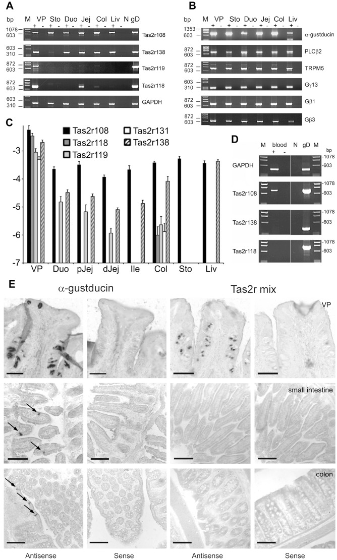
Figure 1. Expression analysis of Tas2rs and taste signaling cascade elements in mouse tissues.
(A) and (B) PCR analyses of cDNA from vallate papillae (VP), stomach (Sto), duodenum (Duo), jejunum (Jej), colon (Col) and liver (Liv). (A) PCR products obtained from cDNA of mouse GI tissues (+) and -RT controls (−) with primers specific for Tas2r108, -r138, -r119 and -r118. GAPDH was amplified as quality control, mouse genomic DNA served as positive control (gD). N, negative control (H2O). (B) PCR analysis of α-gustducin, PLCβ2, TRPM5, Gγ13, Gβ1 and Gβ3 expression in mouse GI tract. M - Molecular weight standard (ΦX174 DNA/HaeIII). (C) Quantitative RT-PCR of gustatory and gastrointestinal tissues. cDNA of vallate papillae (VP), duodenum (Duo), proximal (pJej) and distal (dJej) jejunum, ileum (Ile), colon (Col), stomach (Sto) and liver (Liv) was subjected to quantitative real-time PCR analyses using primers specific for Tas2r108 (black bars), -r118 (dark gray bars), -r119 (light gray bars), -r131 (white bars), and –r138 (hatched bars). Y-axis = logarithm of the expression levels relative to β-actin (mean log(2−ΔCT) ±SE, n≥3). (D) Analysis of Tas2r gene expression in whole blood. PCR products obtained from cDNA of mouse whole blood (+) and -RT controls (−) with primers specific for Tas2r108, -r138, and -r118. GAPDH was amplified as quality control, mouse genomic DNA served as positive control (gD). N, negative control (H2O). M - Molecular weight standard (ΦX174 DNA/HaeIII). (E) In situ hybridization. Antisense riboprobes specific for α-gustducin and a mixture of riboprobes for Tas2r108, -r119, -r138, -r131 and -r118 in VP sections resulted in specific signals in taste buds. In situ hybridization of small intestine sections revealed cells positive for α-gustducin, whereas no signals were detected for Tas2r probes. In situ hybridization in colon sections resulted again in the detection of α-gustducin positive cells, but no signals were evident using Tas2r probes. Hybridization with corresponding sense riboprobes (negative control) showed no staining. Scale bars, 100 µm.

