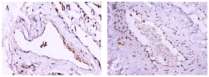
Figure 3. TUNEL assay of middle pulmonary vasculature.

The positive cells were stained in yellow brown by TUNEL assay. Photomicrographs of TUNEL stained lung tissue of the non-COPD group (panel A) and the COPD group (panel B) (magnification: 400×). The number of TUNEL positive pulmonary vascular endothelial cells in the COPD group was much higher than that in the non-COPD group.
