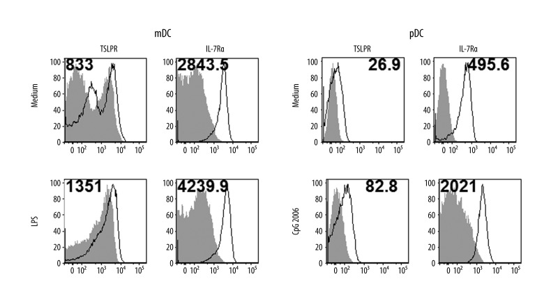
Figure 1.
Expression of TSLPR receptor in dendritic cells. Surface TSLPR and IL-7R expression in myeloid DCs and plasmacytoid DCs was measured by flow cytometry. Cell populations were magnetically separated and in vitro stimulated 24 hours with selected TLR agonists (LPS – lipopolysaccharide, CpG 2006). Open histograms represent staining of TSLPR or IL-7R, shaded histograms represent isotype controls. Mean fluorescence intensity (MFI) of samples substrated with MFI of isotype controls is indicated. Data represent 1 of 3 independent experiments.

