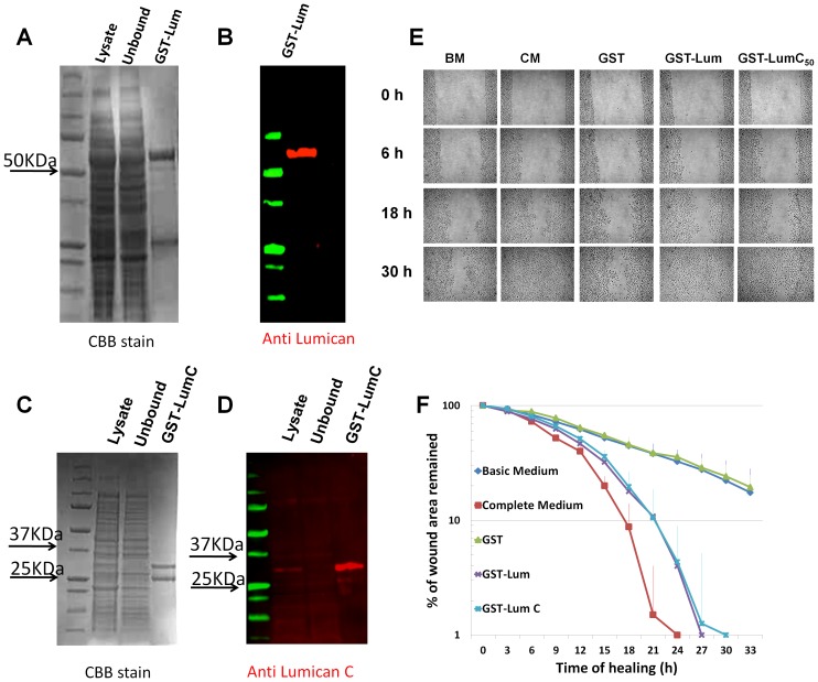
Figure 1. Purification of recombinant Lumican and the healing of scratched HTCE cells.
Purification of GST-Lum and GST-LumC50 recombinant proteins Purification of recombinant GST-Lum and GST-LumC50 was monitored by Coomassie Brilliant Blue (CBB) staining and western blot anlaysis. (A) CBB staining revealed two major bands eluted with glutathione. The upper band with a Mr. ∼70 kDa was GST-Lum and the lower band with a Mr. ∼25 kDa was GST. (B) Immunostaining with an anti-LumN oilgopeptide antibody (CDDLKLKSVPMVPPGIK) only labeled the 70 kDa band (GST-Lum fusion protein) while the lower band did not react to the antibody and is likely related to GST. (C) CBB stained two bands at 30 and 25 kDa from E.coli transfected with GST-LumC50 plasmid. (D) Immunostaining with an anti-LumC peptide antibody (NPLTQSSLPPDMYEC) labeled the 30 kDa GST-LumC50 fusion protein. Effect of recombinant GST-Lum and GST-LumC50 on healing of scratched HTCE cells Confluent HTCE cells were wounded in CM (complete medium), BM (basic medium), BM + GST (glutathione S-transferase recombinant protein), BM+recombinant GST-Lum (0.15 µM) and GSTLumC50. The wound gap was determined by time-lapse microscopy. (E) Representative time-lapse images of the healing of scratched HTCE cells; (F) The healing followed biphasic kinetics in cells treated with BM+GST-Lum and CM, whereas those of BM and BM+GST followed monophasic kinetics. R2 values were as follows: BM 0.957; CM 0.994; GST 0.985; LumC50 0.985; Lum 0.995. The rate constants are summarized in Table 1.

