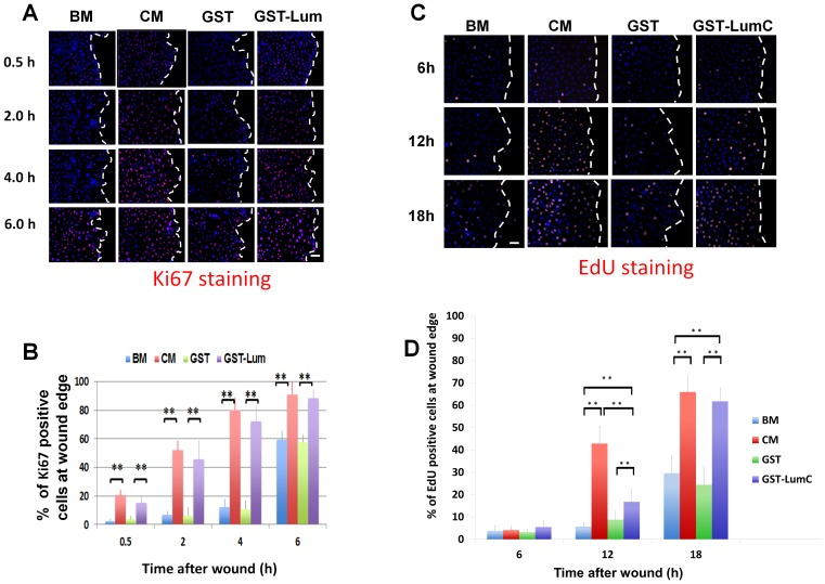
Figure 4. Administration of Lum lifts cell cycle suppression at the wound edge.
Expression of Ki67, a marker of cells engaged in the cell cycle, was determined by immunostaining with an anti-Ki67 antibody at various time periods following wounding. (A) Images of Ki67 expression pattern Treatment with BM or BM+GST contained few Ki67 positive cells at the wound edge 0.5, 2 and 4 h after wounding but showed an increase in expression at 6 h. Treatment with CM and GST-Lum resulted in many Ki67 positive cells at all time points examined. Scale bar = 50 µm. (B) Graphical representation of the percentage of Ki67 positive cells at the wound edge (average±std, n = 12). (C) Representative images of EdU-labeled cells (cells in S-phase) at the wound edge. Few cells at the wound edge were EdU positive at 6 h and 12 h under the treatment of BM and BM+GST, while many EdU positive cells were seen in cells treated with CM and GST-LumC. Scale bar = 50 µm (D) Graphical representation of the percentage of EdU positive cells at the wound edge (mean±std, n = 4). No significant difference was seen at the 6 h time point. More cells at the wound edge enter S-phase 12 hours after wounding in CM and GST-Lum than those with BM and GST treatment. Statistical significance was analyzed by ANOVA. P value of<0.05 was considered significant and indicated by double asterisks. Dashed white lines mark the wound edge.

