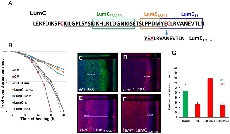
Figure 7. Identification of Lumikines (Lum C-terminal peptide(s) capable of stimulating wound healing in vitro and in vivo.
(A) Amino acid sequences of synthetic LumC peptides, LumC33ΔC20, LumC18ΔC5, LumC13 and LumC13C-A (substitution of C with A). (B) In vitro analysis of the function of LumC peptides on the healing of HTCE cells. Healing followed biphasic kinetics in cells treated with CM, BM+GST-Lum, BM+LumC13 and BM+LumC13C-A; whereas those of BM, BM+LumC33ΔC20 and BM+LumC18ΔC5 followed monophasic kinetics. The R2 values were as follows: full_Lum 0.993; LumC18ΔC5 0.970; LumC33ΔC20 0.936; LumC13 0.952; LumC13c-a 0.902. These data suggest that the last C-terminal 13 amino acids have wound healing activity. (C–G) In vivo wound healing activity of Lumican peptides. To confirm LumC13C-A can also promote corneal epithelium wound healing in vivo, Lum-/- mice were subjected to epithelium debridement. C–F are representative cut view images showing the migration distance. Migration distance was measured between the original wound edge and the wound front. In LumC13C-A treated corneas, the wound front moved ∼144 µm. The control peptide, LumC33ΔC20, did not promote epithelium wound healing. (G) Summary of epithelium migration. The average epithelium migration (µm) and standard deviation were calculated from five corneas of two separate experiments. In PBS and peptide LumC33ΔC20 peptide treated cornea, the epithelium moved ∼38 µm, while corneas treated with LumC13C-A migrated ∼140 µm. Injured corneal epithelium of wild type mice migrated about 100 µm.

