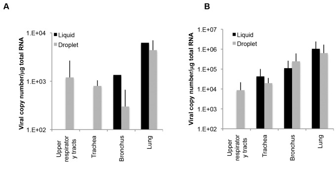
Figure 6. The amounts of viral RNA in the respiratory tract in rhesus and cynomolgus monkeys between different inoculation methods.
(A) A rhesus monkey was inoculated with HPAIV via a catheter. Viral RNA in the respiratory tract (two parts for the upper respiratory tract, three parts for the trachea, two parts for the bronchus, and six parts for the lungs) was quantified by qRT-PCR. The total amount of viral RNA in each organ is indicated as the log value. Data on the three rhesus monkeys (#29−31) shown in Figure 4 was similarly analyzed and the mean±SEM for three monkeys is indicated. (B) Cynomolgus monkeys were inoculated with HPAIV via liquid exposure (n=2) or droplet exposure (n=1). Data were analyzed similarly to (A), including data for cynomolgus monkeys (#23−25). Data are means±SD was indicated.

