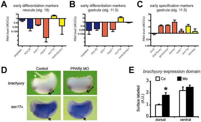
Figure 2. PPARβ promotes differentiation but represses dorsal mesoderm and endoderm specification.
(A)–(C) Embryos were injected with PPARβ MO or Co, allowed to develop until stage 18 (A) or stage 11.5 (B) and (D), and collected for extraction of total RNA. qRT-PCR runs for a selection of neural (blue), mesodermal (red), or endodermal (yellow) markers of differentiation (A) and (B) or of germ layer specification (C) were conducted. RNA levels were normalized to EEF1a and RPL8 and are presented as fold variation between MO and Co samples. Error bars represent the S.E.M. of 3 to 5 independent experiments. (D) Embryos were injected with PPARβ MO or Co, fixed at stg. 11.5, hemi-sectioned along the dorso–ventral axis, and processed for RNA in situ hybridization. While Mo injection did not affect the sox17α expression domain, it resulted in the expansion of brachyury expression dorsally (see the scale) but not ventrally. Arrows indicate the dorsal lip. (E) Quantification of the surface covered by the dorsal and ventral expression domains of brachyury in MO compared to Co hemi-sections. Error bar is the S.E.M. of 10 measurements. *: two-tailed Student’s t-test vs control, P<0.05.

