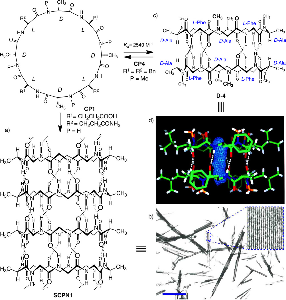
Figure 1.
CP1 and CP4. a) Self-assembled nanotube structure of CP1 (most of the side chains are omitted for clarity). b) Electron microscopy low-magnification image of nanotube suspension adsorbed on a carbon support film (scale bar 1 µm); inset shows low-dose image enhancement of a single crystal and reveals the longitudinal striations that correspond to each nanotube. c) Cylindrical dimer of CP4. d) Crystal structure of D4 emphasizing the internalized water binding sites (blue rod).

