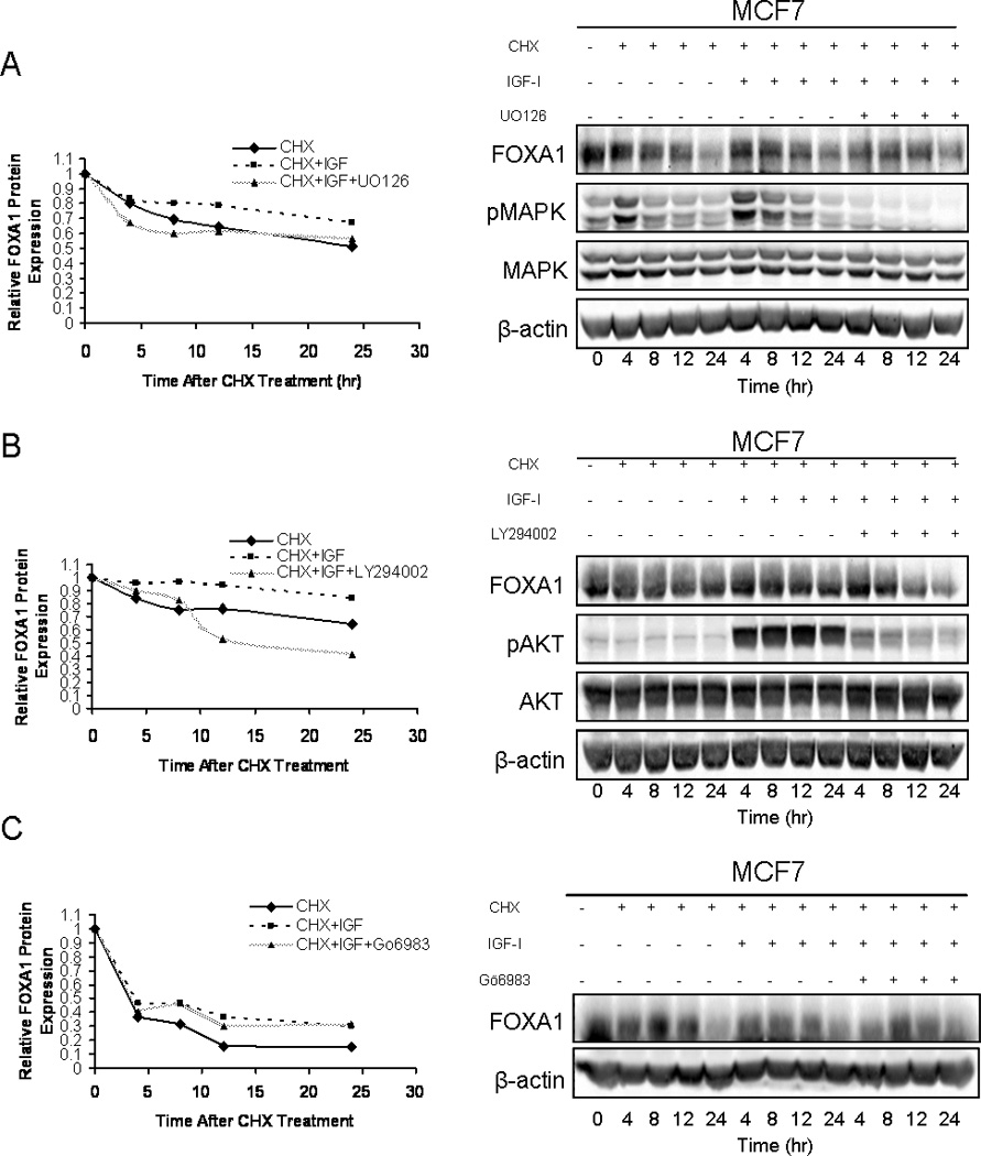
Figure 4. IGF-I increases the half-life of FOXA1 protein by signaling through AKT and ERK 1/2.

MCF7 cells were either treated with cycloheximide (CHX)(20ng/mL), CHX in combination with IGF-I (100ng/mL), or CHX, IGF-I and the MAPK inhibitor U0126 (10µM) (A), PI3K inhibitor LY294002 (20µM) (B), or the PKC inhibitor Gö6983 (0.25µM) (C). Cells were then lysed, protein was harvested and western blot analysis was performed as indicated. Blots represent prototypical examples of experiments replicated at least three times. Quantitative data (OD, optical density) for A, B and C is shown in the left panels.
