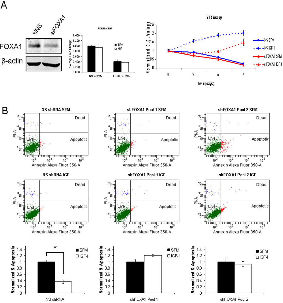
Figure 5. FOXA1 plays a crucial role in IGF-I functional biology.

A) MCF7 cells were transiently transfected with FOXA1 siRNA (red lines) or non-specific siRNA (blue lines) and incubated in either serum-free medium (solid lines) or medium containing IGF-I (100ng/mL) (dashed lines). Cell growth was assessed by MTS assay on days 0, 2, 5 and 7. The assay was performed with biological septuplicates and each point represents the average value ± SEM. B) Cells stably expressing either non-specific shRNA or one of two shRNAs targeted to FOXA1 were cultured in serum-free medium and then either maintained in serum-free medium or treated with IGF-I (100ng/mL) for 4 days. Apoptosis was determined using an Annexin V apoptosis assay. The data are an average of three biological replicates ± SEM. A t-test analysis was performed in which normalized percentage of apoptosis in serum-free conditions was compared to IGF-I treated conditions for non-specific shRNA, shFOXA1 clone1 and shFOXA1 clone2 (* p<0.05).
