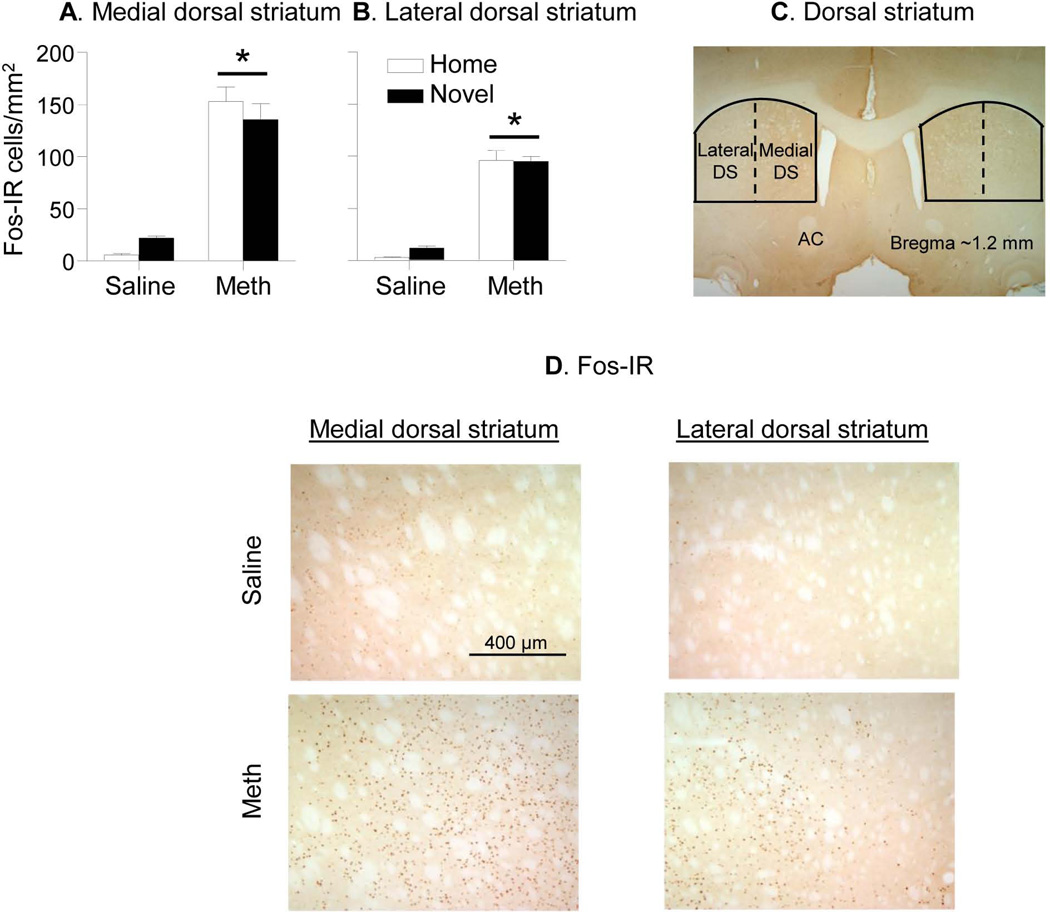
Figure 2. Fos labeling in medial and lateral dorsal striatum of rats injected in Home cage or Novel cage.
(A,B) Number of Fos-IR nuclei per mm2 in medial and lateral dorsal striatum 90 min after Saline or Methamphetamine (Meth) injections. (C) Areas where Fos-IR was quantified. Image was captured using 1.25× objective. (D) Representative images of Fos-IR (dark brown) in the medial (left) and lateral (right) dorsal striatum. Images were captured using 5× objective. Abbreviations: AC: anterior commissure; DS: dorsal striatum. * Different from the Saline condition, p<0.05, n=6 per experimental condition.

