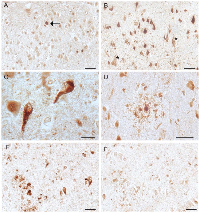
Figure 3.
Immunohistochemistry reveals weak Ob-Rb localization to neuronal cytoplasm in pyramidal neurons of control individuals (A) as well as the occasional age-related neurofibrillary tangles (A, arrow). In the AD cases examined, however, NFT are prominently labeled (B) both intracellullar and extracellular NFT (marked by * in B). The NFT exhibits typical fibrillar morphology (C). Ob-Rb is also colocalized with the neuritic pathology associated with senile plaques (D). Adsorption with the peptide antigen nearly abolishes the NFT and neuritic plaque localization (E) compared to the non-absorbed adjacent sections (F). Scale bars = 50 μm (A,B,D-F) and 20 μm (C).

