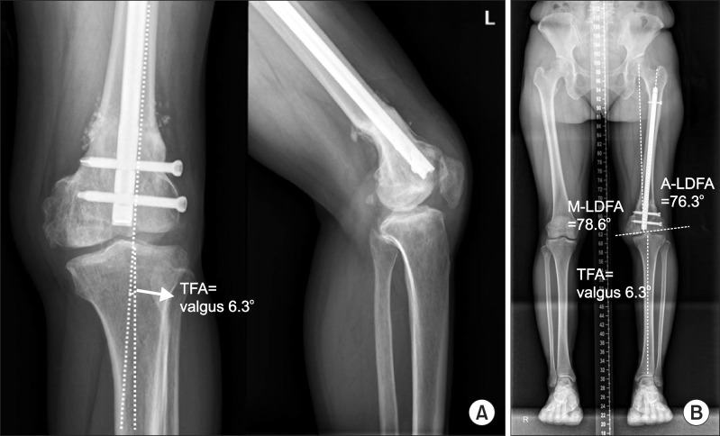
Fig. 5.
Plain radiography after 6 months later deformity correction by supracondylar dome osteotomy with retrograde intramedullary nailing. (A, B) Knee and whole lower extremity plain radiography shows correction of deformity and union of osteotomy site. TFA: tibiofemoral angle, M-LDFA: mechanical lateral distal femoral angle, A-LDFA: anatomical lateral distal femoral angle.

