
Figure 1. Schematic representation of TGF and CTGF.
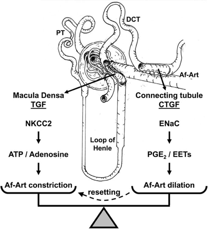
The macula densa triggers TGF when Na is reabsorbed via the Na/K/2Cl cotransporter type 2 (NKCC2), by releasing ATP which is broken down to adenosine, which in turn causes constriction of the Af-Art. The connecting tubule triggers CTGF when Na is reabsorbed via the epithelial sodium channel (ENaC), by releasing epoxyeicosatrienoic acids (EETs) and prostaglandin E2 (PGE2), which cause dilation of the Af-Art. PT indicates proximal tubule, and DCT distal convoluted tubule.