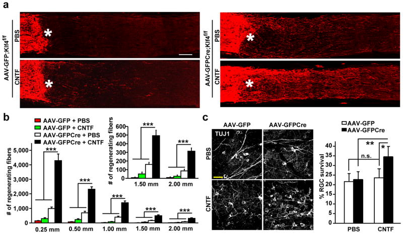
Figure 5. Klf4-deletion enhances cytokine-induced axon repair and survival.
(a) Representative confocal images showing CTB-traced RGC axons 16 days post nerve crush. Asterisks, crush sites; Scale, 100 μm. (b) Estimated axon numbers at the indicated distance to the crush sites (means ± s.e.m.; n = 5; ***p < 0.001 by ANOVA with Bonferroni’s post-test). (c) Survival of TUJ1+ RGCs 16 days post injury. Confocal images of TUJ+ RGCs are shown in the left panels. The number of TUJ1+ RGCs in the injured retina was normalized to that in the un-injured retina within the same animal (means ± s.e.m.; n = 5; *p < 0.003 and **p < 0.001 by ANOVA with post-hoc Tukey’s test; n.s., not significant).

