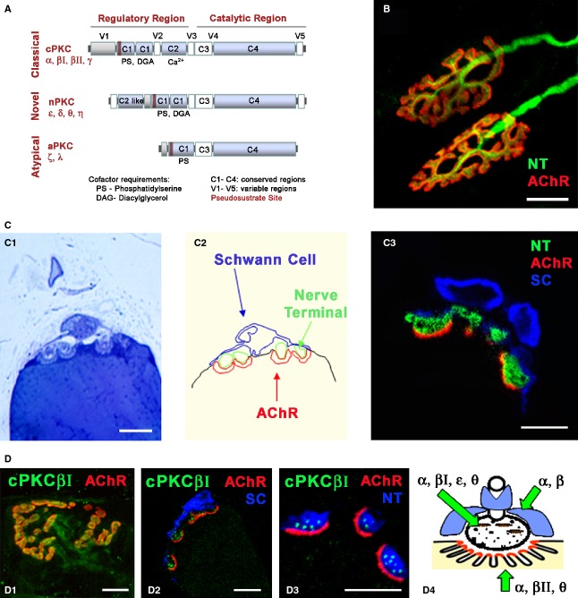
Fig. 1.
Protein kinase C and NMJ. (A) Serine/threonine kinase C family. Schematic diagram of primary structures of PKC family members showing domain composition and activators. The PKC family of isozymes consists of three classes: the classical (α, βI, βII, γ), novel (δ, ε, η and θ) and atypical (ζ and ι/λ). (B) Adult NMJ image. Double immunofluorescence NMJs labelled with syntaxin/neurofilament-200 nerve terminal, NT, in green) and α-BTX (AChR in red). (C1–3) Semithin (0.5 μm) cross-sections of the NMJ stained with toluidine blue (C1) and with a triple immunofluorescence method (C3; syntaxin/neurofilament-200-NT, in green, S-100 Schwann cell, SC, in blue and α-BTX -AChR in red). In (C2), the cellular components in C1 are delineated. Reproduced with permission from Lanuza et al. (2007). (D) Immunohistochemical staining for cPKC isoform βI at the adult NMJ. cPKCβI are labelled in green, the AChRs in red and the Schwann cells (SC, S-100) or the nerve terminals (NT, neurofilament-200/syntaxin) in blue. (D1) NMJ from a whole muscle immunolabelled. (D2–D3) Semithin cross-sections from a whole-mount multiple-immunofluorescent stained muscle. cPKCβI was found to be localized to the presynaptic terminals. Reproduced with permission from Besalduch et al. (2010). (D4) The diagram summarizes the localization of the PKC isoforms in the three cellular components of the NMJ (nerve terminal, muscle cell and Schwann cell). Scale bars: 10 μm.

