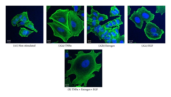
Figure 1.

Combined stimulation by TNFα + Estrogen + EGF induces extensive morphological changes and spreading in breast tumor cells. Breast tumor cells were either (A1) not stimulated (cells grown in the presence of diluents) or stimulated by (A2a) TNFα (50 ng/mL), (A2b) estrogen (10−8 M), (A2c) EGF (30 ng/mL), or (B) TNFα + Estrogen + EGF (concentrations as above) for three days. The stimulatory conditions were selected following titration and kinetics analyses (data not shown). Actin filaments were detected by phalloidin staining (green) and cell nuclei by DAPI staining (blue). The cells were analyzed by confocal microscopy. In all panels, the results are from a representative experiment of n ≥ 3.
