Figure 10.

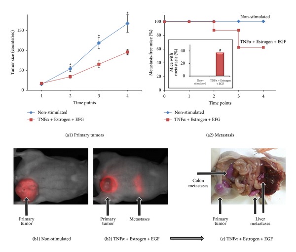
In response to combined stimulation by TNFα + Estrogen + EGF, breast tumor cells acquire high metastasizing abilities. mCherry-expressing breast tumor cells were stimulated by TNFα + Estrogen + EGF (concentrations as in Figure 1) for three days. Non-stimulated: cells grown with the diluents of the above factors. Following washing, equal numbers of live cells (4 × 10⁶) were inoculated to the mammary fat pad of mice. Using the CRi Maestro intravital imaging system, tumors and metastases were followed in intact mice at four different time points along the experiments, up to 37 days. (a) Followup of primary tumors in the mammary fat pad and formation of macrometastases. (a1) Sizes of tumors at the mammary fat pads are presented as counts/sec of fluorescence emission, divided by 1,000, obtained at each time point by analyses with the CRi Maestro intravital imaging system. *P < 0.05 for differences between the two groups of mice. The figure sums up the results obtained in two experimental repeats showing similar results, with a total n = 6 mice in the control group n = 8 mice in the group of mice inoculated with cells stimulated with TNFα + Estrogen + EGF. (a2) Kaplan-Meier analyses of metastasis-free mice, showing incidence of macrometastases detected by the Maestro device in intact animals in four time points along the experiments, up to 37 days. The figure sums up the results obtained in two experimental repeats showing similar results, with a total of n = 6 mice in the control group and n = 8 mice in the group of mice inoculated with cells stimulated with TNFα + Estrogen + EGF. Inset: the incidence of mice with macrometastases at the end-point of the experiments, determined in intact mice by the Maestro device (38% in the TNFα + Estrogen + EGF-stimulated tumor cells versus 0% in the control group, in two independent experiments providing similar results). # Macrometastases were also observed in 2/3 mice in another experiment of TNFα + Estrogen + EGF cells (in which control mice were not included). (b) Representative pictures obtained by the Maestro device in intact mice, showing tumor cells (red, mCherry) in both groups of mice. Non-stimulated tumor cells (b1) developed bigger tumors than tumor cells stimulated with TNFα + Estrogen + EGF (b2); however, macrometastases were detected only in the group of mice administered with tumor cells stimulated by TNFα + Estrogen + EGF (the image was obtained following prolonged excitation of mCherry in the CRi Maestro, in order to visualize the metastases). (c) A representative picture of the macrometastases that have developed in mice inoculated with tumor cells stimulated by TNFα + Estrogen + EGF, at the end of the experiment. The image shows the same mouse demonstrated in part (b2). Because of the expression of mCherry, the tumor cells carried a purple color. In this representative mouse, metastases were detected in the liver, colon, and above the kidney.
