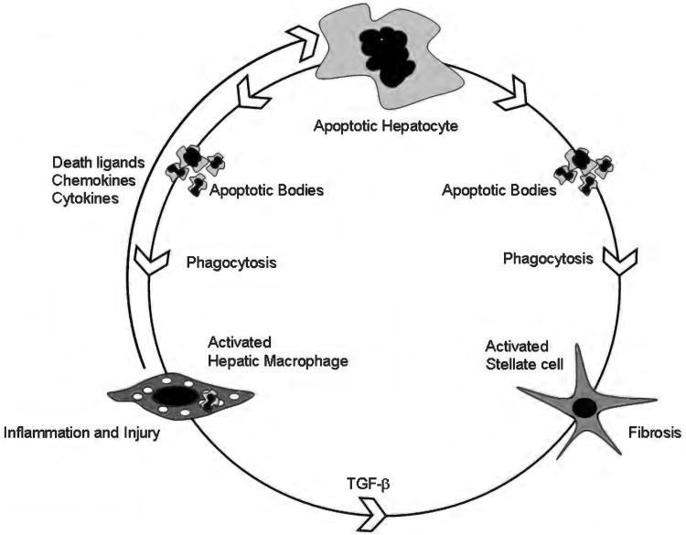
Figure 2.
Apoptosis-inflammation-fibrosis in the liver. This cartoon depicts the circular relationship between apoptosis, inflammation, and fibrosis in the liver. Hepatocyte apoptosis is the central event in the model shown. In the setting of an apoptotic stimulus, for example, toxic bile salts or palmitic acid, a vulnerable hepatocyte undergoes cell death. Apoptotic bodies are formed. These can be engulfed both by hepatic macrophages, also known as Kupffer cells and hepatic stellate cells (54, 57). Macrophages, upon activation, in turn release death ligands, such as Fas ligand and TRAIL, both of which can induce hepatocyte apoptosis. Inflammatory cytokines such as TNF-α, IL-1β, and IL-6 are also released by activated macrophages (54). These result in liver inflammation and injury. Engulfment of apoptotic bodies in a permissive milieu (inflamed liver with increased fibrogenic signals, such as Transforming growth factor (TGF)-β) results in activation of hepatic stellate cells to myofibroblasts (57). These cells remodel the extracellular matrix resulting in fibrosis and cirrhosis.

