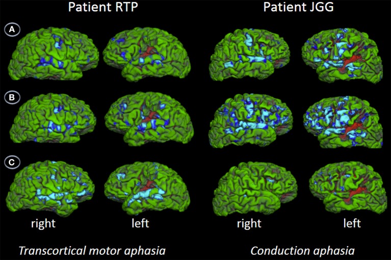
Figure 5.
Functional MRI. The figures show the contrast words (A), nonwords (B), and word triplets (C) versus rest. Contrasts are shown on patients' uninflated cortical surface of the right and left hemispheres and significant activations (p < 0.05, corrected) are depicted in light blue and blue. The fMRI shows bilateral perisylvian activation in both patients in all three tasks. Although JGG obtained lower performance in word and nonword repetition tasks than RTP, he showed greater areas of activation, extending into motor, premotor and prefrontal areas in addition to the perisylvian areas activated by both of them. In contrast, word triplet repetition activated a greater bilateral network in RTP than JGG. See further details in text and Table 3.

