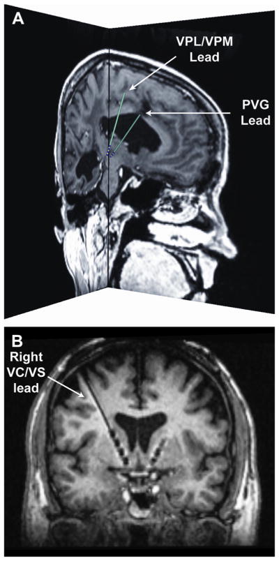
Fig. 1.

DBS targets for pain management. (A) More traditional DBS targets aimed at treating the sensory-discriminative component of pain. The image shows the preoperative magnetic resonance imaging (MRI) and corresponding lead (model 3387, Medtronic, Minneapolis, MN) locations for a patient with one DBS lead in the VPL/VPM and a second DBS lead in the PVG. Both sagittal and coronal slices are shown near the distal ends of the leads. The patient-specific lead locations and trajectories were determined using the software Cicerone v1.3.25 (B) A recently proposed DBS target aimed at treating the affective-motivational component of pain.24 The image shows an oblique coronal view of the postoperative MRI for a patient with bilateral DBS leads (Medtronic model 3391) implanted in the ventral capsule and ventral striatal (VC/VS) area. It is possible to see the 4 electrodes in the right hemisphere as well as the distal end of the DBS lead implanted in the left hemisphere.
