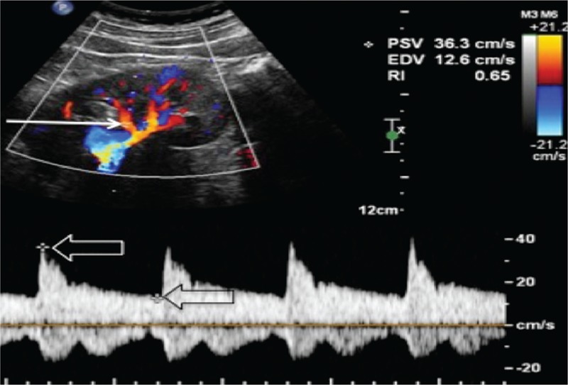
FIGURE 1.

Renal RI measurement technique. A sample volume (arrow) is placed within an intrarenal artery (an arcuate or interlobar one) under Color Doppler guidance and spectral analysis of vascular signals is obtained. The measurement calipers are then set at the systolic peak (white open arrow) and end diastole (black open arrow) of a waveform, and the RI is calculated according to the formula (PSV-EDV)/PSV. EDV, end diastolic velocity; RI, resistive index; SV, peak systolic velocity.
