Figure 5.
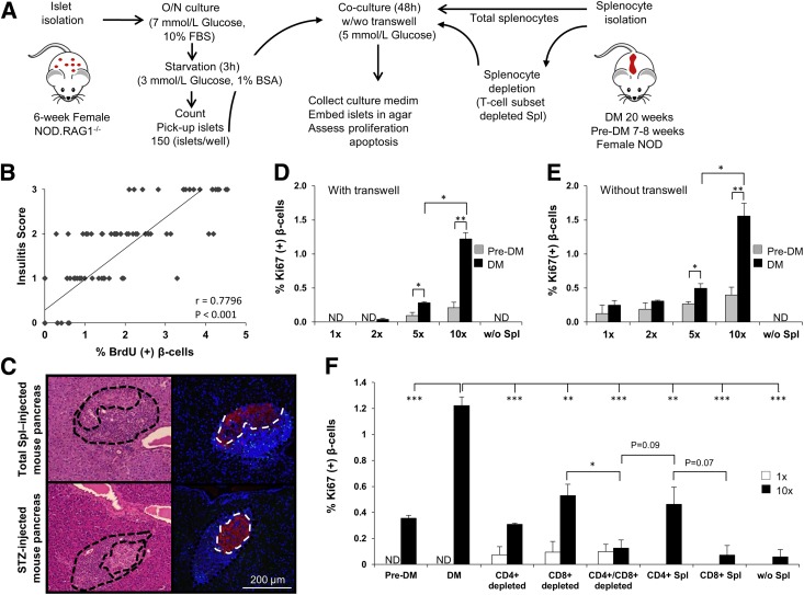
β-cell proliferation is stimulated by infiltrating lymphocytes via soluble factors. A: Experimental strategy showing total splenocytes (DM or pre-DM) or depleted splenocytes (diabetic mice), cocultured with 5–6-week-old NOD.RAG1−/− mouse islets (150 islets/condition) for 48 h in 5 mmol/L glucose in the presence or absence of transwell inserts. B: Linear regression of insulitis score and BrdU+ β-cells in single pancreatic islets harvested from NOD.RAG1−/− mice transferred with total diabetic splenocytes. Each square represents a single islet out of 120 analyzed islets. r = 0.80; P < 0.001. C: Representing pancreatic sections derived from total splenocyte– or STZ-injected mice used for determining the ratio between infiltrating immune cells vs. insulin+ β-cells. Islets are indicated by dotted lines in the right panel. The area of infiltration is shown around the islet in the left panels. β-cell proliferation in agar-embedded NOD.RAG1−/− islets cocultured with total splenocytes from DM or pre-DM at a ratio of 1:1, 1:2, 1:5, or 1:10 or without splenocytes in the presence (D) or absence (E) of transwell inserts (n = 3–4). *P < 0.05; **P < 0.01 (Student t test). F: Quantification of proliferating β-cells in pancreatic islets cocultured with total (DM or pre-DM), negatively, or positively selected DM splenocytes at 1:1 or 1:10 ratio with transwell conditions (n = 3–6 for each condition). *P < 0.05; **P < 0.01; ***P < 0.001 (Student t test). Data are expressed as means ± SEM. ND, not detected; O/N, overnight; Spl, splenocyte.

