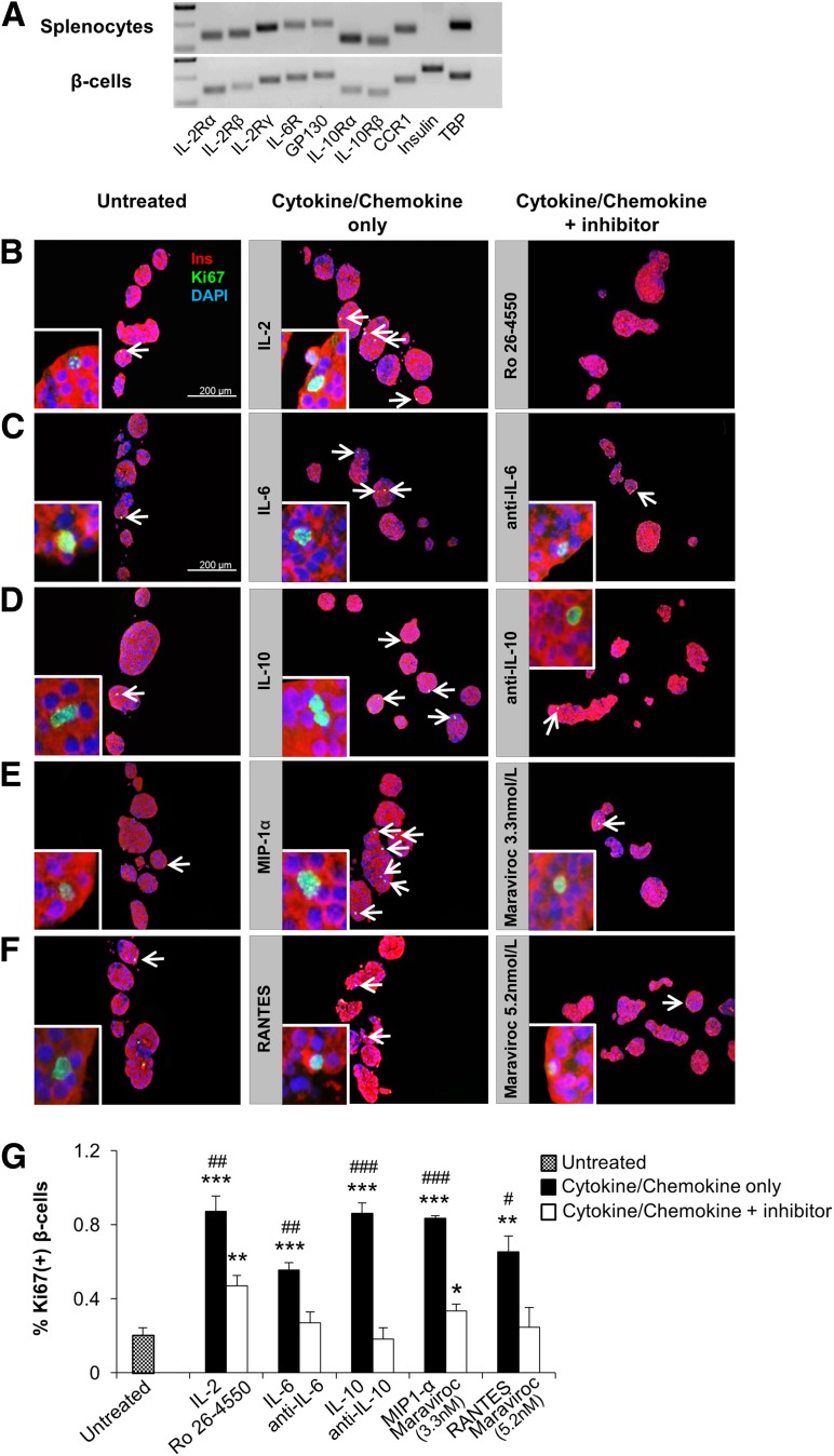
Figure 7.
Effect of soluble factors on β-cell proliferation is reversed by inhibitory/neutralizing antibody treatment in vitro. A: Detection of the cytokine/chemokine receptor subunit mRNAs by real-time PCR from sorted β-cells and splenocytes harvested from C57BL/6 mice. Tata-box-binding protein (TBP) was used as reference. B–F: Agar-embedded pancreatic islets from C57BL/BJ mouse treated in the absence (control) or presence of low-dose recombinant proteins with or without inhibitory/neutralizing molecules (as described in research design and methods) for 48 h (150 islets/condition, three to four replicates). Representative sections are shown. Islets were costained for the proliferation marker Ki67 (green) with insulin (red) and DAPI (blue). Arrows indicate proliferating β-cells (Ki67+/insulin+). Scale bar, 200 µm. Insets show a magnified image of a representative proliferating β-cell. G: Quantification of data in B–F (n = 3–4 in each group). *, #P < 0.05; **, ##P < 0.01; ***, ###P < 0.001 (Student t test). *, untreated vs. cytokine/chemokine or inhibitory/neutralizing antibody treated; #, cytokine/chemokine treated vs. inhibitory/neutralizing antibody treated. Data are expressed as means ± SEM. CCR1, C-C chemokine receptor type 1; GP130, glycoprotein 130; IL-2R, IL-2 receptor.

