Figure 8.
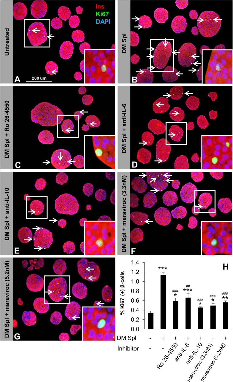
Lymphocyte-secreted soluble factors enhance β-cell proliferation in vitro. One hundred fifty agar-embedded islets harvested from C57BL/6 mice cocultured without (untreated control) (A) or with total DM splenocytes in the absence (treated control) (B) or presence of inhibitory/neutralizing molecule Ro 26-4550 (C), anti–IL-6 (D), anti–IL-10 (E), maraviroc 3.3 nmol/L (F), or maraviroc 5.2 nmol/L (G). Islets were costained for the proliferation marker Ki67 (green), insulin (red), and DAPI (blue). Arrows indicate proliferating β-cells (Ki67+/insulin+). Scale bar, 200 µm. Insets show magnified image of a representative proliferating β-cell. H: Quantification of proliferating β-cells in A–G (n = 3–4 in each group). *P < 0.05; **, ##P < 0.01; ***, ###P < 0.001 (Student t test). *, untreated vs. DM splenocyte treated; #, DM splenocyte treated vs. inhibitory/neutralizing antibody treated. Data are expressed as means ± SEM. Ins, insulin; Spl, splenocyte.

