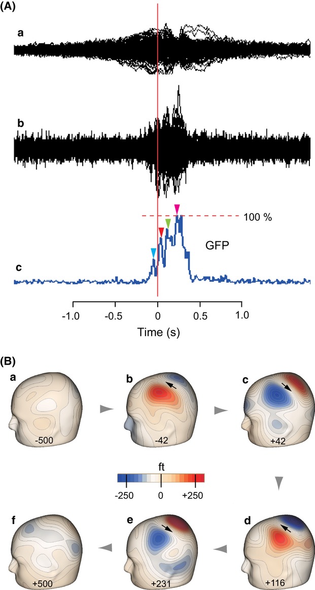
Figure 1.

Movement-related cerebral fields following pulsatile extension of the index finger. Data from a representative subject. (A) Superimposed waveforms of all the channels without (a) and with low cut filtering (b). For the latter, the corresponding global field power (GFP) curve is shown in c, in which prominent peaks for specifying motor field (MF) and MEFI–III components are indicated by blue, red, green, and magenta arrowheads, respectively. (B) Six snapshots (a–f) of isocontour maps of evoked magnetic fields in the left hemisphere contralateral to the movement at chosen peaks are indicated by arrowheads in the GFP curve in A-c. The negative values in panels a-b and positive values in panels c-f indicate latency before (−) and after (+) the movement, respectively. Arrows in panels b–e show the location and orientation of estimated equivalent current dipoles (ECDs).
