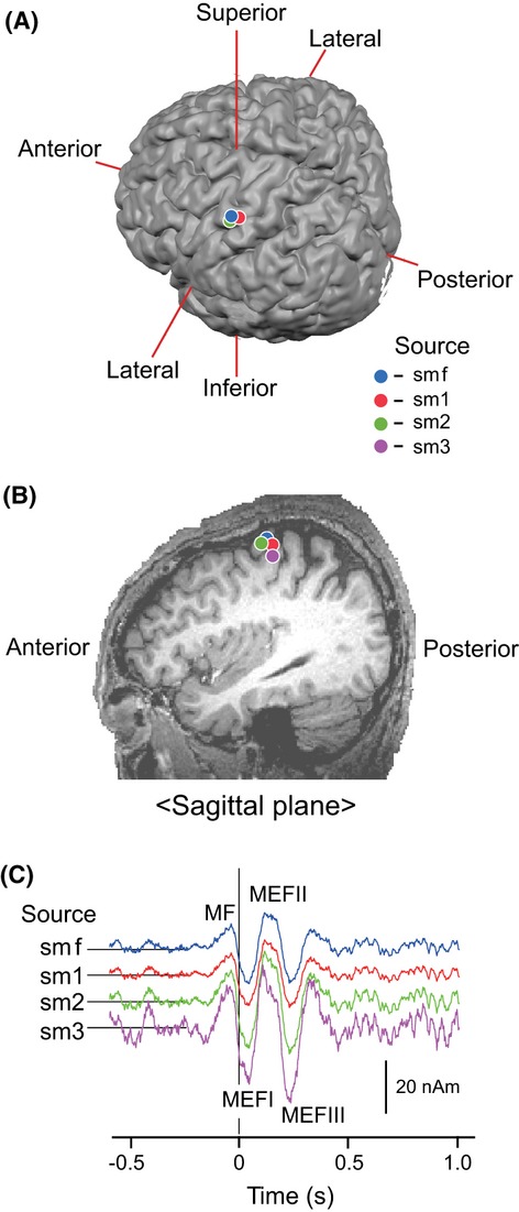
Figure 2.

Spatiotemporal characteristics of source response modeled for movement-related cerebral fields (MRCFs). (A) Superpositions of four dipole sources (smf, sm1–sm3) on an MR image in posterior/superior oblique view. (B) The same superpositions of four dipoles sources in the sagittal plane view through the motor cortex region in the left hemisphere. Note that three or four plots seem to locate in nearly the same position in the posterior crown (smf, sm1, and sm2) in A or the wall of the precentral gyrus (smf, sm1–sm3) in B. (C) Comparison of the time course of source strength among four different dipole sources, smf (blue), sm1 (red), sm2 (green), and sm3 (magenta).
