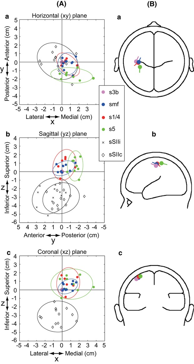
Figure 6.

Simultaneous representation for spatial locations and orientations of four independent sources in somatosensory evoked fields (SEFs) and smf in MRCFs. (A) Plots for locations of all sources in SEFs (i.e., s1/4, s5, sSIIi, and sSIIc) and of smf are presented relative to the positions of s3b (defined as the cross-point of vertical and horizontal lines in the figure) in three orthogonal planes; from top to bottom, horizontal (a), sagittal (b), and coronal (c) planes. In each panel, an ellipse represents a 95% confidence limit (z-value: 1.96) for corresponding plots of source location in each of three orthogonal planes. (B) Grand-averaged source locations and orientations of four independent sources in SEFs and smf. The same sources to those depicted in A are averaged and depicted. In each source, the orientation of the current is represented by the line segment embedded in the plot for the location of the corresponding source. In three anatomical planes, source orientations are represented as their projection on three orthogonal planes, that is, horizontal (xy), sagittal (xz), and coronal (yz) plane, in panels a to c, respectively.
