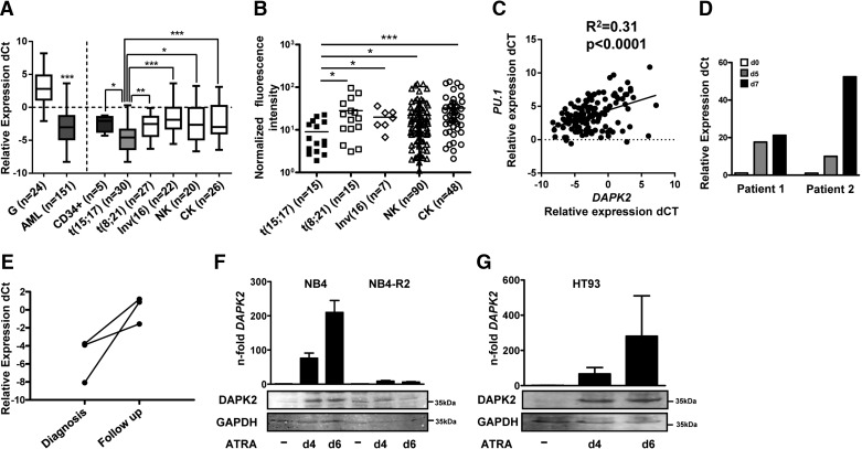
Figure 1. DAPK2 is down-regulated in AML.
(A) DAPK2 mRNA levels in AML blasts from the HOVON/SAKK/Bern cohort in granulocytes from healthy donors and in CD34+ progenitor cells were quantified using qPCR. The relative mRNA expression levels were given as differences in comparative threshold (dCt) values compared with mRNA levels for the housekeeping genes GAPDH and ABL1. Values are the differences in Ct values between DAPK2 and the housekeeping genes and ABL1 and GAPDH. ABL1=Abelson tyrosine-protein kinase 1, CK=complex karyotype, G=granulocytes, NK=normal karyotype. (B) DAPK2 mRNA expression in AML patient samples from the Munich cohort was measured by microarray hybridization. (C) Positive correlation between DAPK2 and PU.1 mRNA levels in the HOVON/SAKK/Bern AML patient cohorts (n=133), as measured by qPCR. (D) DAPK2 mRNA levels during a short-term follow-up of two newly diagnosed APL patients treated with ATRA were evaluated. Values are normalized to the housekeeping genes ABL1 and HMBS and are given as n-fold increase relative to Day (d) 0 of treatment. (E) DAPK2 mRNA levels in a long-term follow-up of three APL patients receiving ATRA (45 mg/m2) daily for up to 60 days. Values are the differences in Ct values between DAPK2 and the housekeeping genes ABL1 and HMBS. (F and G) DAPK2 is up-regulated during neutrophil differentiation of APL cells. DAPK2 mRNA and protein levels in NB4, HT93, and ATRA-resistant NB4-R2 APL cells were measured by qPCR and Western blotting, respectively. mRNA levels were normalized to the HMBS housekeeping gene and the nontreated control cells. Results of at least three independent experiments are shown as n-fold regulation to control cells. Total protein was extracted and submitted to immunoblotting using anti-DAPK2. GAPDH is shown as a loading control. MWU: *P < 0.05; **P < 0.01; ***P < 0.001.

