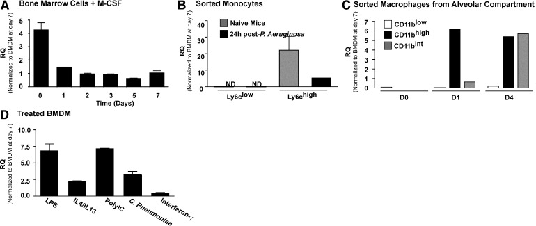
Figure 1. Expression of Mmp28 in myeloid cells.
(A) Expression of Mmp28 in BM cells at time of harvest and during differentiation to macrophages in the presence of M-CSF relative to expression at Day 7 (set to one). Mmp28 expression decreased during culture, with a slight increase at Day 7. Overall, expression remained significant at all times (n=3/genotype/time-point). RQ (Fold increase). (B) Expression of Mmp28 by monocyte populations in naive (gray bars) or infected (black bars) mice reveals selective expression of Mmp28 by Ly6chigh monocytes, with reduced expression after infection (n=3 mice/genotype). (C) Expression of Mmp28 in macrophage subpopulations isolated from the alveolar space reveals greater expression of Mmp28 by recruited macrophages (CD11bint, CD11bhigh) compared with resident cells (CD11blow; n=3 mice/genotype). (D) Expression of Mmp28 in stimulated BMDM (relative to Day 7, untreated BMDM). LPS, IL-4/13, Poly(I:C), and C. pneumoniae all increased Mmp28 gene expression, with the exception of IFN-γ, which did not alter Mmp28 expression significantly (n=3–6/genotype/condition).

