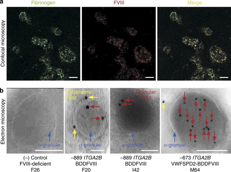
Figure 2. Synthesis and trafficking of BDDFVIII into canine platelet α-granules.
(a) Confocal microscopy showing co-localization of BDDFVIII and Fg within platelets. Canine CD34+G-PBC were transduced with lentivirions encoding human BDDFVIII followed by transplant haemophilia A dogs. Peripheral blood platelets were isolated from whole blood, fixed, permeabilized and examined by indirect immunofluorescence analysis for Fg and BDDFVIII distribution. Shown is a representative image using a × 10 eye piece and × 100 oil objective and a × 2 digital zoom of platelets isolated from one transplanted animal (I42) from an experiment that was performed seven times on all three dogs and a FVIII-deficient negative control. Fg was visualized with a 1°Ab to this platelet-specific marker for α-granules and a Alexa488-conjugated 2°Ab (left, green). BDDFVIII was detected with 1°Ab to human FVIII and an Alexa568-conjugated 2°Ab (middle, red). BDDFVIII colocalized with Fg is observed when the two images are merged (right, yellow). The white scale bar is 5 μm in length. (b) Electron microscopy localized human BDDFVIII directly in α-granules. Shown is a representative image of BDDFVIII absent from an ultrathin cryosection of a single platelet α-granule (blue arrow) from a FVIII-deficient (negative control, F26) when probed with a 1°Ab (301.3) to human BDDFVIII and a 2°Ab-conjugated to 10-nm gold particles. In contrast, BDDFVIII was localized directly within the α-granule (red arrow) and within intracytoplasmic membrane systems (yellow arrow) in representative images from all three experimental dogs (F20, I42 and M64). Interestingly, sections from dog (M64) receiving the −673ITGA2B-VWFSPD2-BDDFVIII-transduced G-PBC (VWF-targeting peptide) exhibited a noticably higher concentration of BDDFVIII within mature platelet α-granules. Each sample was cut on a minimum of three separate occasions with a series of ultrathin sections subjected to immunogold labelling and analysis of at least 100 α-granules per sample. The white scale bar is 0.2 μm in length.

