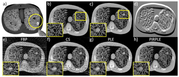
Figure 11.

CBCT phantom experiments using a) an anthropomorphic chest phantom with natural sponges simulating lung tissue and a 0.5 inch removal acrylic sphere to simulate a lung nodule. Fully sampled CBCT acquisitions (450 mAs) in two scenarios (no nodule vs. nodule) we collected to reconstruct a) a prior image (no nodule); and b) a fully sampled reference image (with nodule). Rigid motion of the phantom between the two data acquisitions was induced and is apparent in the difference image in d). Sparse current data acquisitions (4.9 mAs) in the second scenario were used to produce reconstructions e–h) using FBP, CS, PLE, and PIRPLE.
