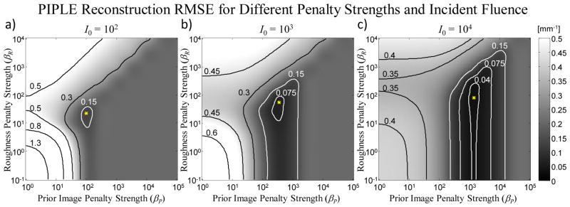
Figure 4.

RMSE in reconstructions of the lung nodule image [Figure 2(b)] from sparse projections over a range of prior image penalty strengths (βP) and roughness penalty strengths (βR) for three levels of incident fluence: (a) 102, (b) 103, and (c) 104 photons / pixel. In this case, δ is fixed at 10−4 mm−1. The optimum (minimum RMSE) is marked with an asterisk.
