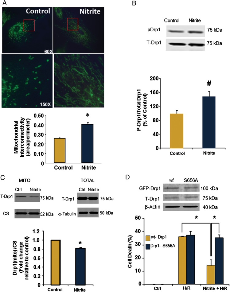
Figure 2.
Nitrite promotes mitochondrial fusion by increasing Drp1 phosphorylation. Cells were treated with or without nitrite (25 µmol/L; 21% O2, 30 min) and collected 1 h after nitrite removal. (A) Cells stained with antibodies to TOM20. Top panel is ×60 magnification and bottom panel is ×150 magnification of image section in red box. Quantification of interconnectivity using n = 20–23 fields/group. (B) Representative immunoblot for phosphorylated and total Drp1 and quantification of Phospho-Drp1 levels (normalized to total Drp1). n = 4. (C) Representative immunoblot and quantification of Drp1 expression (and citrate synthase; CS) in whole cells and the mitochondrial fraction of cells treated with or without nitrite (25 µmol/L). (D) Protein expression of exogenous (GFP-Drp1) and endogenous (T-Drp1) in transfected cells. Cell death after H/R in wild-type (yellow) or cells expressing the S656A mutant (blue) treated with or without nitrite. Asterisks indicate P < 0.01 and hash indicates P < 0.05 vs. control; n = 5 per group.

