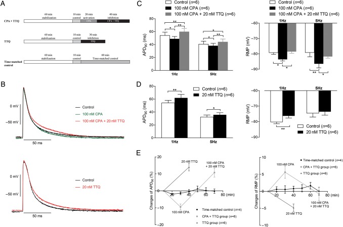
Figure 4.
Effects of GIRK channel activation and inhibition on isolated papillary muscle. (A) Perfusion protocols. In the CPA+TTQ group the protocol comprised the following periods: 60 min of stabilization, 10 min of baseline recording, 20 min of GIRK channels activation by 100 nM CPA, followed by 40 min of GIRK channel inhibition by 20 nM TTQ in the presence of 100 nM CPA. In the TTQ group, the protocol consisted of 60 min of stabilization, 10 min control recordings, and then 30 min perfusion with 20 nM TTQ in the absence of GPCR activation. In the time-matched control group, tissues were perfused for 70 min after stabilization. (B) Representative recording from the CPA + TTQ group (upper panel) and the TTQ group (lower panel), tissue paced at 1 Hz. (C) Effect of CPA and TTQ on APD90 (left panel) and RMP (right panel) in the CPA + TTQ group (n = 6). (D) Effect of TTQ on APD90 (left panel) and RMP (right panel) in the TTQ group (n = 6). (E) Changes of APD90 (in %, left panel) and RMP (in %, right panel) in the time-matched control group (n = 4) show that APD90 and RMP were stable throughout the whole experiment. *P < 0.05, **P < 0.01.

