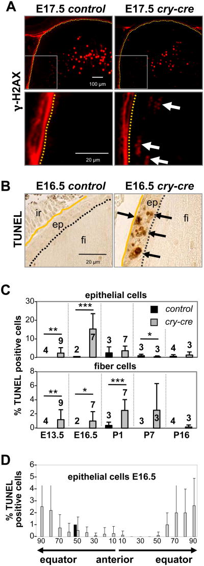
Fig. 4. Loss of E2F1-3 causes increased cell death without affecting differentiation.
(A) Epithelial cells of E17.5 E2f1-/-;E2f2-/-;E2f3LoxP/LoxP (control) and cry-cre;E2f1-/-;E2f2-/-;E2f3LoxP/LoxP (cry-cre) lenses stained with antibodies for γ-phosphorylated H2AX, a form of H2AX protein recruited to DNA double-strand breaks. Note that H2AX- positivity is present during normal degradation of nuclear contents, required for organelle degradation and maturation of lens fiber cells. Aberrant positivity was observed near the bow region of the mutant lenses. (B) TUNEL staining at E16.5 shows apoptosis in the lens. ep, epithelium; fi, fiber cells; ir, iris. (C) Quantification of the percentage of TUNEL-positive cells shows elevated levels of cell death in the epithelium and fiber compartment. A minimum of three sections near the central plane were analyzed for cry-cre lenses (grey bars) at E13.5 (n=9), E16.5 (n=7), P1 (n=7), P7 (n=3), and P16 (n=3) and for control lenses (black bars) at E13.5 (n=4), E16.5 (n=2), P1 (n=3), P7 (n=3), and P16 (n=4). ep, epithelium; fi, fiber cells; ir, iris. Error bars represent standard deviation. Significance of unpaired T-test indicated by * p < 0.05, ** p < 0.01, and *** p < 0.001. (D) Spatial distribution of TUNEL-positive cells in control lenses (black bars) and cry-cre lenses (grey bars); note the increased apoptosis near the transition zones of cry-cre lens equators.

