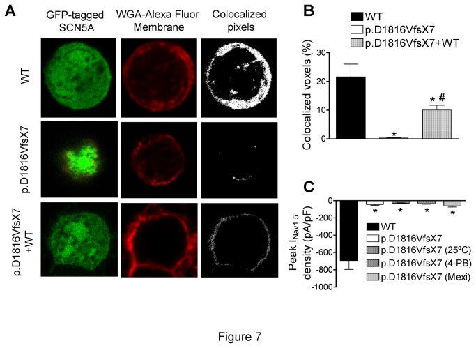
Figure 7. WT and p.D1816VfsX7 Nav1.5 channel trafficking.
A. Representative single fluorescent confocal images from the center of CHO cells transfected with GFP-tagged-Nav1.5 WT, p.D1816VfsX7, and p.D1816VfsX7+WT constructs. Columns: GFP-tagged-Nav1.5 channel staining (left), wheat germ agglutinin Alexa Fluor 647 (a fluorescent membrane dye) staining (middle), and colocalization of both (right). B. Percentage of colocalized voxels measured in cells transfected with WT, p.D1816VfsX7, and p.D1816VfsX7+WT. C. Comparison of peak current-density recorded in cells expressing p.D1816VfsX7 incubated or not at 25°C, or in the presence of 5 mM 4-phenylbutirate (4-PB) or 300 μM mexiletine (mexi) with WT. Bars represent the mean±SEM of >6 cells. * P<0.05 vs WT and # P<0.05 vs p.D1816VfsX7.

