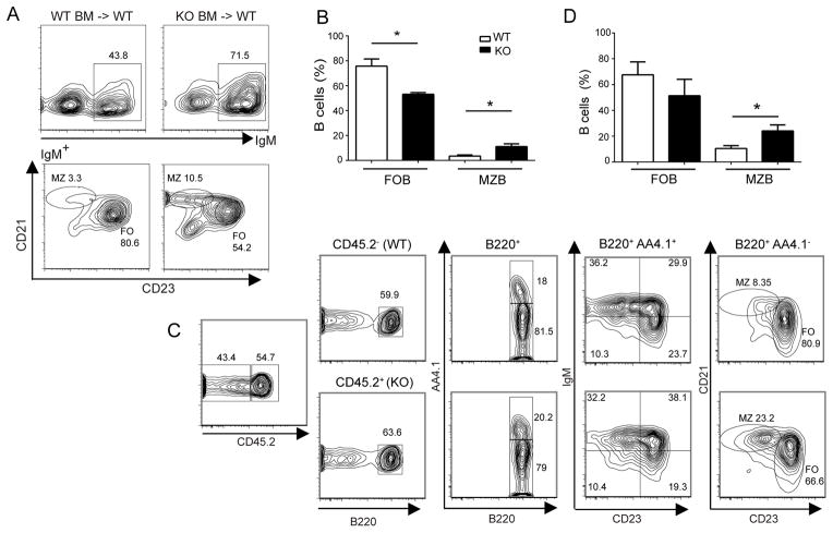
Figure 2.
Increase in MZ B and decrease in FO B cells in Bcl-3−/− mice is due to a B cell autonomous defect. (A) Lethally irradiated WT mice were reconstituted via transfer of bone marrow cells from WT or KO mice. Representative flow cytometric analysis of splenocytes stained for expression of surface markers with gates across top of panels as indicated. (B) Summary graph of data as in (A) shown as the mean ±SEM from 2 separate experiments with n=5 independently analyzed recipient mice. (C) Irradiated Rag1−/−mice were reconstituted via co-transfer of approximately equal numbers of bone marrow cells from CD45.1+ (CD45.2−) WT and CD45.2+ KO mice. Representative flow cytometric analysis of splenocytes stained for expression of surface markers with gates as indicated. (D) Summary graph of data as in (C) shown as the mean ±SEM from 3 separate experiments with n≥4 independently analyzed recipient mice. Abbreviations as in Fig. 1. (*= p<0.05)

