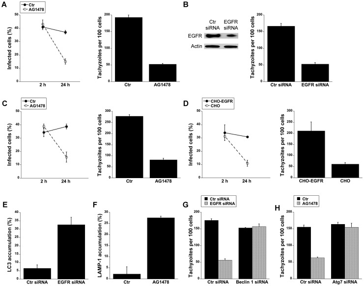
Figure 6. Blockade of EGFR induces accumulation of the autophagy protein LC3 around the parasite, vacuole-lysosome fusion and killing of T. gondii dependent on the autophagy proteins.
A, HBMEC were incubated with AG1478 (1 µM) 1 h prior to challenge with T. gondii. Monolayers were examined by light microscopy at 2 and 24 h. B, Human RPE cells transfected with either EGFR or control siRNA were challenged with T. gondii followed by examination of monolayers by light microscopy at 24 h. C, Mouse bone marrow-derived macrophages were incubated with AG1478 and challenged with T. gondii. Monolayers were examined by light microscopy at 2 and 24 h. D, Parental CHO and CHO-EGFR cells were challenged with RH T. gondii. Monolayers were examined microscopically 2 h or 24 h post-challenge. E, mHEVc-LC3-EGFP cells transfected with control siRNA or EGFR siRNA were challenged with T. gondii-RFP. Monolayers were examined by fluorescence microscopy 5 h post-challenge to determine the percentage of endothelial cells with LC3 accumulation around the parasite. F, HBMEC cells treated with or without AG1478 were challenged with either T. gondii-YFP. Expression of LAMP-1 was examined by fluorescent microscopy 8 h post-challenge. The percentages of endothelial cells with LAMP-1 accumulation of around the parasite were determined. G, H, mHEVc cells transfected with Beclin1 siRNA (G) or Atg7 siRNA (H) were transfected with EGFR siRNA or treated with or without AG1478 followed by challenge with T. gondii. Monolayers were examined by light microscopy 24 h post-challenge. Results are shown as the mean ± SEM and are representative of 3 independent experiments.

