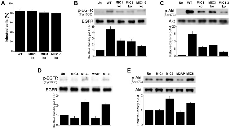
Figure 8. T. gondii micronemal proteins appear to induce EGFR and Akt activation.
A, B, C HBMEC were challenged with ΔHx (WT), MIC1 ko, MIC3 ko, MIC1-3 ko T. gondii at MOIs that yielded similar percentages of infected cells (A). Cell lysates were obtained and used to examine total EGFR and phospho-tyrosine 1068 EGFR (B) or total Akt and phospho-Ser 473 Akt (C) by immunoblot. HBMEC were incubated with 10 nM of recombinant MIC4, MIC3, M2AP or MIC6 for 15 minutes followed by examination of total EGFR and phospho-tyrosine 1068 EGFR (D) or total Akt and phospho-Ser473 (E) by immunoblot. Densitometry data represent means ± SEM of 4 experiments. Band densities from infected cells or cells treated with MICs were compared to bands from uninfected or untreated cells (Un), which were given an arbitrary number of 1. Results shown are representative of 4 independent experiments.

