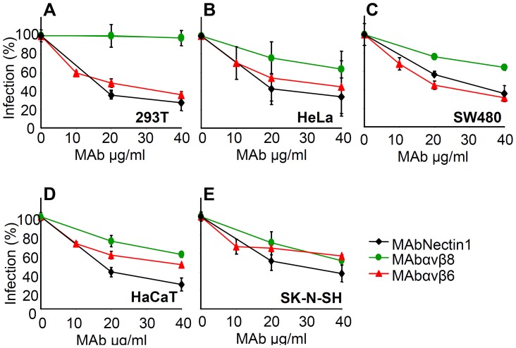
Figure 3. Inhibition of HSV-1 infection by function blocking MAbs to αvβ6- or αvβ8–integrin.
293T (A), HeLa (B), SW480 (C), HaCaT (D), SK-N-SH (E) cells were exposed to increasing amounts of MAb to nectin1 (black diamond), to αvβ6-integrin (red triangle), to αvβ8- integrin (green circle) for 1 h, infected with R8102 (3 pfu/cell) in the same medium, and overlaid with MAb-containing mediun until harvest at 6–8 h after infection. The extent of infection was quantified from Lac-Z gene engineered in the viral genome under the immediate-early α27-promoter. Extent of β-Gal activity, measured as conversion of ONPG substrate, reflects the amount of infection. Each point represents the average of triplicates. ONPG conversion was measured by O.D. reading at 405 nm [5]. 100% infection is the value obtained with murin IgG used as a control. Bars show SD.

