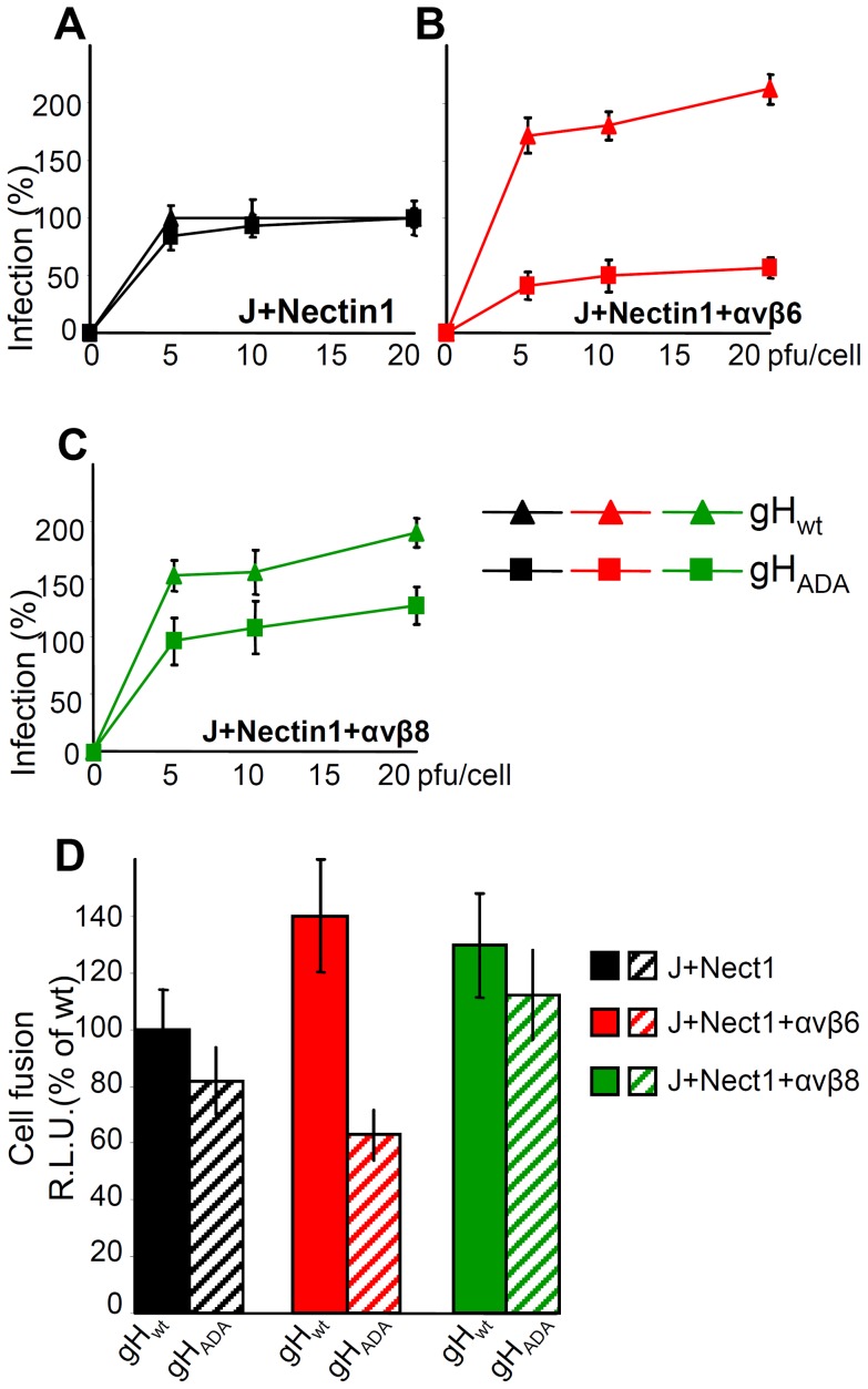
Figure 8. The RGD motif in gH is required for HSV infection and cell-cell fusion mediated by αvβ6–integrin, but not by αvβ8–integrin.
(A–C). J cells were transfected with low amount of nectin1 alone (black triangle and square) (A), or nectin1 plus αvβ6-integrin (red triangle and square) (B), or nectin1 plus αvβ8-integrin (green triangle and square) (C), as detailed in legend to Fig. 7. 48 h later, cells were infected with increasing amounts (5–20 pfu/cell) of SCgHZ virus, a ΔgH HSV, complemented with gHwt (black, red and green triangle) or with gHADA (black, red and green square), and harvested 16–18 h later. Extent of infection was expressed as detailed in legend to Fig. 3. Eeach point represents the average of triplicates. 100% of infection is the value obtained in J cells transfected with nectin1 alone and infected with 20 pfu of SCgHZ virus complemented with gHwt. Bars show SD. (D) Cell-to-cell fusion between effector J cells expressing gHwt, or gHADA plus the trio of gD, gL and gB, and luciferase, and target J cells expressing nectin1 alone, or nectin1 plus αvβ6-integrin, or nectin1 plus αvβ8-integrin plus Renilla luciferase. Fusion was quantified by means of a T7 promoter-driven reporter luciferase gene transfected in effector cells, and expressed as percentage relative luciferase units (R.L.U.). 100% is the value obtained in cells expressing nectin1 alone and gHwt plus the trio of gD, gL, gB. Each point represents the average of triplicates. Bars show SD.

