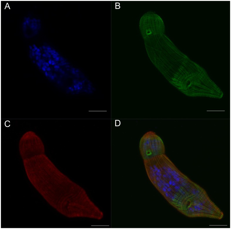
Figure 4. Septin fibers co-localize with actin fibers at the surface of the schistosomulum.
Panel A: Cross-section at the surface of a two day old schistosomulum showing DAPI stained nuclei. B: F-actin structure stained with phalloidin. C: Septin labeled with anti-SmSEPT5. D: Merged channels. Scale bars, 20 µm.

