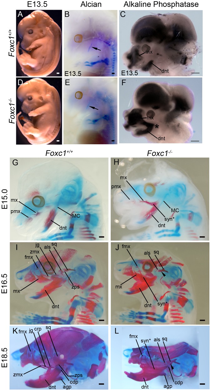
Figure 6. Time-course of PA1-derived skeletal abnormalities in Foxc1−/− mice.
(A, D) Gross view of fixed Foxc1+/+ (A) and Foxc1−/− (D) embryos at E13.5. Cerebral hemispheres are enlarged and the snout is foreshortened in the mutant. (B, E) Whole mount alcian blue staining to detect cartilage differentiation in the wild type (B) and mutant (E) embryos pictured in (A, D). A single, normal Meckel's cartilage (arrows in B, E) is seen in both control and Foxc1−/−. (C, F) Bissected E13.5 heads of Foxc1+/+ (C) and Foxc1−/− (F) embryos stained for endogenous alkaline phosphatase (AP) activity to detect early osteoblast differentiation. In the absence of Foxc1, early osteoblasts of the dentary (dnt) and maxillary (mx) region initially differentiate in a fused, syngnathic pattern (*). (G–L) Whole mount alcian blue (cartilage) and alizarin red (bone) staining of Foxc1+/+ (G, I, K) and Foxc1−/− (H, J, L) embryos. (G, H) At E15.0, ossification of the wild type maxilla (mx) with a wispy frontal process (fmx) and dentary can be seen. In the mutant, wispy projections are seen in the maxillary region similar to controls. However, this ossification is connected to a larger ossified region that is fused (syn*) directly to the dentary. (I–L) In Foxc1+/+ embryos at E16.5 (I) and E18.5 (K), the zygomatic process of the maxilla (zmx), jugal (jg), zygomatic process of the squamosal (zps), and alisphenoid (als) are clearly identified as separately ossifying elements. By contrast, in the Foxc1−/− specimens (J, L), syngnathia is observed in the zygomatic region, rather than the distinct elements found in wild type. The alisphenoid and the squamosal (sq) are hypoplastic compared to controls. The zygomatic process of the squamosal does not form (arrowheads). Scale bars: 500 µm Abbreviations: agp, angular process; cdp, condylar process; crp, coronoid process; mx, premaxilla; tongue, tg.

