Figure 4. Between P7 and P10 the OBNS/PC were infected with lentivirus transducing GFP.

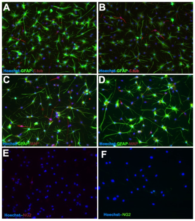
Differentiation of OBNS/PC-GPP was applied between passage 12-15. The differentiated OBNS/PC-GFP exhibited positive immunoreactivity for GFAP astrocytes marker (60-70%) (Figure 5, A,B,C,D), MAP2 mature neuronal marker (15%) (C, D), β-TubulinIII immature neuronal marker (8%) (A,B), NG2 positive cells w/o T3 + PDGFAA (E) (7-8%), and with T3 + PDGFAA (F) (16%). The nuclei were stained blue with Hoechst.
