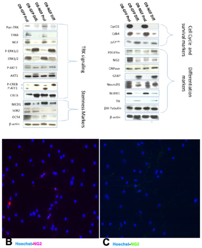
Figure 6. Western blot data revealed that the NGF protein levels look similar although the NGF levels were higher in differentiated OBNS/PC-GFP-NGF respect to OBNS/PC-GFP.

Up- regulation of early oligodendroglia markers (PDGFRα, NG2 and CNPase) was observed in OBNS/PC-GFP-hNGF respect to OBNS/PC-GFP. Sox2 and Oct4 were up-regulated in OBNS/PC-GFP vs. OBNS/PC-GFP-hNGF. Nurr1, TH, NeuroD1 and β-TubulinIII expression were similarly expressed in the 4 cell populations. Survival genes (ERK, AKT1, CREB) were differently modulated between the 4 classes of cell populations; high-affinity Trk receptor was down modulated in differentiated cell populations vs proliferated ones. B. Without T3+PDGFAA, 14% of the differentiated OBNS/PC-GFP-hNGF exhibited positive immunoreactivity for NG2 oligodendrocyte marker. C. In the presence of T3+PDGFAA, almost 25% of differentiated OBNS/PC-GFP-hNGF were NG2 positive. The nuclei were stained blue with Hoechst.
