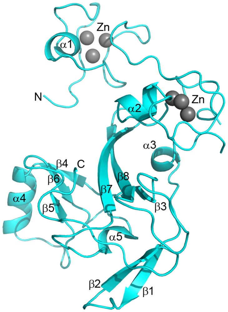
Figure 2. The crystal structure of the EZH2-SET domain.
The crystal structure of the EZH2-SET domain is represented as a ribbon model colored cyan. Bound zinc atoms are represented by spheres and colored gray. Secondary structure elements are labeled. The crystal structure contains two N-terminal zinc binding domains each of which binds three zinc molecules. The core of the domain is formed by β-strands 3, 7, and 8. This core is flanked on one side by a three stranded antiparallel β-sheet (β-4, β-6, β-5) and an accessory α-helix (α-4) and bounded below by α-5, β-1, and β-2. The C-terminus turns upward insinuating through the substrate binding cleft between the β-5/β-6 loop and β-7.

