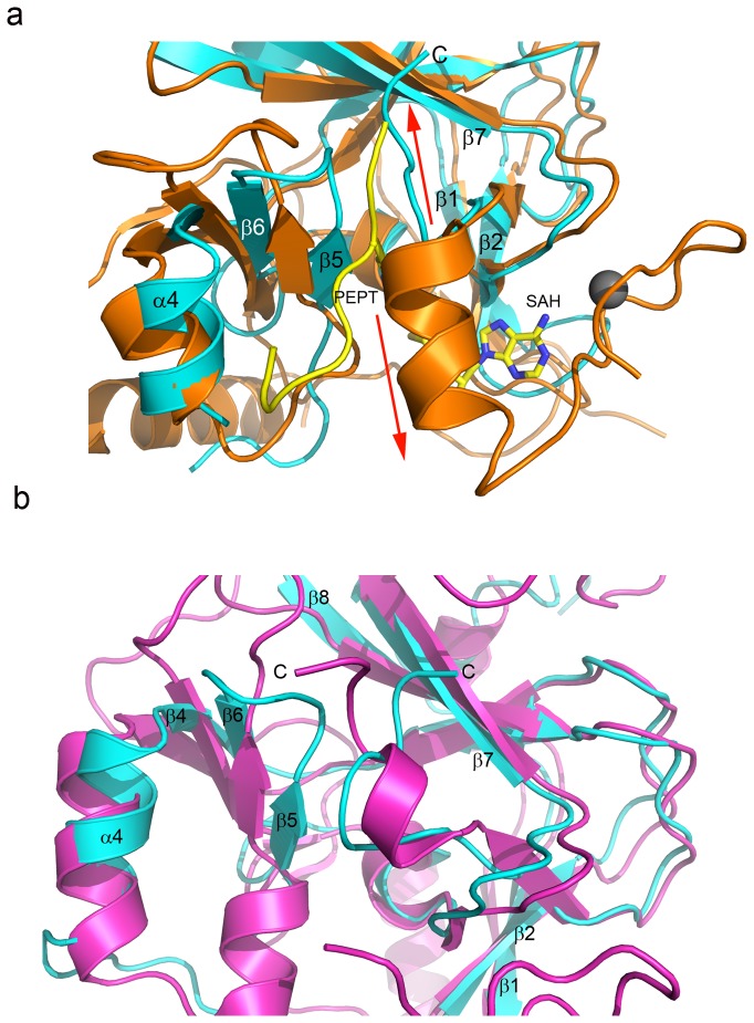
Figure 3. The EZH2-SET domain C-terminus partially occupies the substrate binding groove.
(a) The EZH2-SET (cyan) and hEHMT1-SET (orange) (PDB ID:3HNA) domains are superimposed and represented by ribbons. Zinc bound by hEHMT-SET is represented as a gray sphere. The substrate peptide bound by hEHMT1 is a yellow ribbon with the lysine side chain represented as sticks. The SAH bound by hEHMT1-SET is represented by sticks and colored by atom (carbon, yellow; oxygen, red; nitrogen, blue; sulfur, sienna). The C-terminal tail of EZH2-SET turns upwards and occupies the upper region of the substrate binding groove (red arrow pointing up). The C-terminus of hEHMT1-SET turns downward (red arrow pointing downward) forming the lower lobe of the cofactor binding pocket and coordinating one zinc atom. (b) The EZH2-SET (cyan) and SUV39H2 SET domain (magenta) (PDB ID:2R3A) crystal structures are superimposed and represented by ribbons. The C-termini in both structures occupy the collapsed substrate binding groove.

