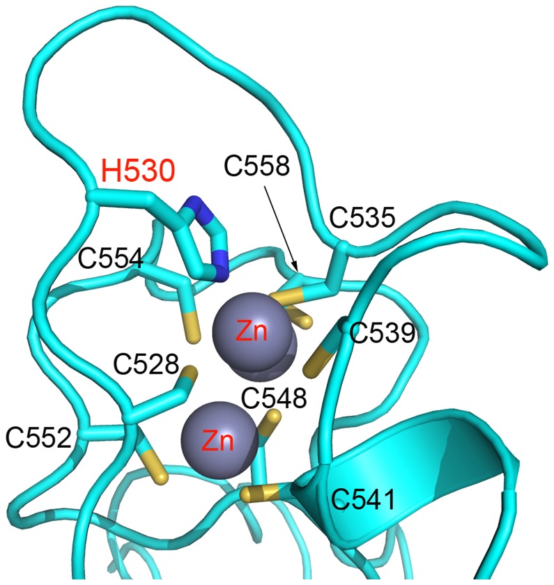
Figure 7. Location of mutation in the first zinc binding domain of EZH2-SET.
EZH2-SET (cyan) is represented as a ribbon diagram with zinc atoms shown as gray spheres and side chain represented as sticks (carbon, cyan; nitrogen, blue; sulfur, sienna) A H530N mutation was identified in AML [29]. This mutation disrupts coordination of zinc in the first zinc binding domain likely having a strong destabilizing effect on the protein.

